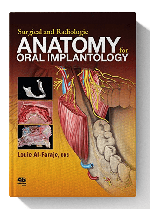
Anatomical textbooks and atlases often fail to meet the clinical demands of defining intraoperative structures for oral implantologists because of the overwhelmingly detailed minutia.
Because certain anatomical landmarks are hard to illustrate in a diagram format, students and professionals can be confused when confronted with actual specimens in the dissecting room or in the operatory.
This book, however, shows the structures of the maxilla, the mandible, and the nasal cavity as they actually exist in the dissected or live body, through the presentation of cadaver specimens and clinical cases.
Several of the chapters include full-page images of specific cadaver sections with all the relevant anatomical parts labeled for convenience.
Cone beam computed tomography images are also presented to show how this technology can be used to measure the bone density, the width of the alveolar ridge, and the exact distance available for implant placement under or above certain anatomical landmarks prior to implant selection.
This book will simplify the learning and execution of implant-related surgical procedures in a region of the body that presents special topographic and anatomical difficulties.
Frequently Asked Questions
Which payment methods are accepted?
We accept all major credit/debit cards, PayPal, Apple Pay, and Google Pay. All transactions are processed through our PCI‑compliant gateway for maximum security.
Can I read the book on my phone or tablet?
Absolutely. Any PDF‑capable reader—such as the free Adobe Acrobat app, Apple Books, or Google Drive viewer—will open the file. Zooming and bookmarks are fully supported
Do I need special software to open the PDF?
No special software is required—any standard PDF viewer works. We recommend the latest version of Adobe Acrobat Reader (free) for the best experience.






Reviews
There are no reviews yet